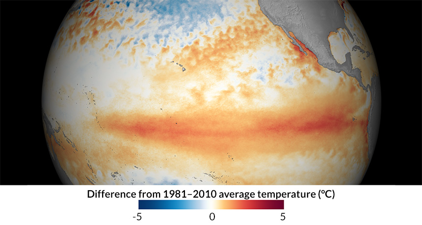
The historic El Niño event currently shaking up Earth’s weather rose like a phoenix from the hot remains of a failed 2014 El Niño, new research suggests.
In 2014, the scientific community buzzed about the possibility of a supersized El Niño as warm Pacific Ocean water sloshed eastward. That July, however, large winds pushed westward and halted the budding El Niño before it fully formed (SN: 11/1/14, p. 6). Those same winds also prevented the release of stored-up ocean heat, researchers report in a paper to be published in Geophysical Research Letters. In March 2015, that lingering heat gave the current El Niño a jump start toward the extreme, the researchers propose. The ongoing El Niño is among the three strongest on record (SN Online: 7/16/15); it has boosted rainfall in California, contributed to ocean coral bleaching and helped make 2015 the hottest year on record (SN: 2/20/16, p. 13). Such a once-in-a-generation El Niño would have been less likely without the failed 2014 event, says study coauthor Michael McPhaden, a physical oceanographer at the National Oceanic and Atmospheric Administration’s Pacific Marine Environmental Laboratory in Seattle.
“In a sense, we dodged a bullet in 2014 by not having a monster El Niño,” McPhaden says. “But that was short-lived, because the conditions that shut that developing El Niño down set up the big one in 2015.”
El Niños typically form every two to seven years when Pacific winds shift a large, near-surface pool of warm water eastward. That warm water then rises to the surface and releases its heat into the atmosphere, causing global shifts in storms, precipitation and temperatures.
The fizzled 2014 El Niño followed by a colossal event in 2015 is very unusual, McPhaden says. He and climate scientist Aaron Levine, also at NOAA’s Pacific Marine Environmental Laboratory, wondered if the sequence of events was just a coincidence. So the researchers looked at decades of El Niño climate data and ran computer simulations of various hypothetical El Niño events.
Under typical ocean conditions, the chances of a 2015 El Niño of any strength are about 27 percent, the researchers estimate. The remnant heat from the failed 2014 El Niño increased those odds to roughly 40 percent. Having a failed El Niño the previous year stacks the deck in favor of an El Niño, McPhaden and Levin conclude. But it “isn’t a guarantee,” Levine says. A similar aborted El Niño occurred in 1990, the researchers find. An El Niño formed the following year, but the event ended up being more modest than the current super El Niño. That’s in part because the eastward-blowing winds in 1991 were relatively weak, Levine says. Strong El Niños require strong winds, not just warm water, he adds.
Forecasting those winds is tricky because the winds and the warm water “are all part of the same system,” says Kevin Trenberth, a climate scientist at the National Center for Atmospheric Research in Boulder, Colo. Ocean heat can cause atmospheric changes that can in turn influence the winds. The new work provides insights, he says, “but it is far from complete.”
Packing on a few pounds may not be such a bad thing.
As a group, overweight people are living the longest nowadays, suggests an almost four-decade study in Denmark published May 10 in JAMA. And obese people seem to be at no higher risk of dying than those of normal weight. The new analysis fuels ongoing debate about what’s a healthy body mass index — especially in light of rising obesity rates (SN: 5/14/16, p. 5), improved heart health treatments and other factors influencing health and longevity. “This is a very carefully done study,” says Rexford Ahima, a physician who studies endocrine disorders at the University of Pennsylvania School of Medicine. The findings strengthen the notion that “BMI as a number alone may not be sufficient to predict health and risk of death. It has to be taken within context.” Ahima was not involved in the research but has analyzed previous studies urging a rethink of how BMI influences mortality.
Researchers screen for obesity by calculating BMI — a popular but fairly crude measurement of body fat reached by dividing a person’s weight in kilograms by the square of height in meters. People with BMIs between 18.5 and 24.9 are considered normal. A BMI between 25 and 29.9 is “overweight”; 30 and above is “obese.”
Many studies suggest that obese individuals face a higher risk of heart disease, stroke and other ills. But some analyses have found that heavier folks may not in fact be in such dire straits. In one study of type 2 diabetes patients, those with normal weight when diagnosed were more likely to die than those who were overweight or obese (SN: 9/8/12, p. 13). And a 2013 meta-analysis of 97 studies found that being overweight was associated with lower risk of death than having a normal BMI — a surprising finding that echoed a 2005 study by the same researchers.
In this new analysis, clinical biochemist Børge Nordestgaard of Copenhagen University Hospital and his team studied more than 100,000 adults. The three groups of white Danes, recruited about 15 years apart, reflected the general population in Copenhagen.
From 1976 to 2013, BMI associated with lowest risk of death increased from 23.7 to 27. That falls squarely in the overweight category. What’s more, obese individuals had the same mortality risk as people in the normal range, the analysis found. That trend held even when researchers took into account potentially confounding factors including age, sex, smoking and a history of cardiovascular disease or cancer. While some might misinterpret the study to mean “you can eat as much as you like,” this is not what the findings suggest, Nordestgaard says. Rather, the results indicate that people who are moderately overweight might not need to worry as much as they had in the past. That might be because better treatments are now available for high blood pressure, high cholesterol and other risk factors for heart disease, Nordestgaard speculates. “So maybe you can be overweight if you have [these conditions] treated.” But the study was not designed to address whether improved heart health care actually caused “healthy” BMI values to creep up over time.
It’s also unclear whether the results apply to other ethnic groups. A substantial fraction of Asians, for instance, develop type 2 diabetes and heart disease despite having BMIs lower than the existing cutoff point for being overweight.
The findings underscore the idea that a person’s BMI does not tell the whole story. While this measure is good for comparing populations, it is not as useful for evaluating individuals and their risk for disease and death, Ahima says. Interpreting an individual’s BMI depends on many other factors, including “whether you are man or woman, how much muscle you have, how physically fit you are and what diseases you have.”
Houseflies stretch their legs to land. Bumblebees hover, then slowly descend. Now, insect-sized flying robots have a way to stick the landing, too.
A tiny aerial bot about the size of a bee (nicknamed RoboBee) uses static electricity to cling to the underside of a leaf and perch on other materials, study coauthor Robert Wood of Harvard University and colleagues report in the May 20 Science.
RoboBee, a bot with shiny, flapping wings and four pinlike legs, is the first of its size that can fly, perch on a surface and then take off again. This energy-saving feat could one day extend mission time in search and rescue operations, the researchers say. For robots, tackling the problem of flight has been easier than figuring out how to land. “Engineers have been trying to build perching mechanisms for flying robots nearly as long as we have been creating flying robots,” Wood says. Researchers have had success with bigger, bird-sized bots (SN: 2/7/15, p. 18), but their landing mechanisms are tricky to scale down. For the microbot, Wood and colleagues wanted something simple: lightweight and without moving parts.
The team created an “electroadhesive” patch with electrodes that can be charged, letting the patch stick to different surfaces, like a balloon sticking to the wall after being rubbed on someone’s hair.
Switch the electrodes on and the patch, a circular disc on top of the robot, helps RoboBee hang out on overhanging pieces of glass or plywood, for example. Switch the electrodes off and the bot detaches, free to fly again. The sticky contraption lets RoboBee rest between flights: The bot used about a thousandth as much energy perching than hovering, the researchers found.
Painkillers in the opium family may actually make pain last longer. Morphine treatment after a nerve injury doubled the duration of pain in rats, scientists report the week of May 30 in the Proceedings of the National Academy of Sciences.
The results raise the troubling prospect that in addition to having unpleasant side effects and addictive potential, opioids such as OxyContin and Vicodin could actually extend some types of pain. If a similar effect is found in people, “it suggests that the treatment is actually contributing to the problem,” says study coauthor Peter Grace, a neuroscientist at the University of Colorado Boulder. Scientists have known that opioid-based drugs can cause heightened sensitivity to pain for some people, a condition called opioid-induced hyperalgesia. The new study shows that the effects linger weeks after use of the drugs is stopped. Male rats underwent surgery in which their sciatic nerves, which run down the hind legs, were squeezed with a stitch — a constriction that causes pain afterward. Ten days after surgery, rats received a five-day course of either morphine or saline.
Rats that didn’t receive morphine took about four weeks to start recovering, showing less sensitivity to a poke. Rats that got morphine took about eight weeks to show improvements — double the time. “That’s far bigger than we had anticipated,” Grace says. “We were definitely surprised by that.”
These experiments were done with male rats, but unpublished data indicate that morphine extends pain even longer in female rats, Grace says, results that fit with what’s known about differences in how males and females experience pain.
Longer-lasting pain in the rats came courtesy of an inflammatory response in the spinal cord. The immune system sees morphine as a threat, the researchers suspect, and responds by revving up inflammation through specialized cells called microglia. Experiments that shut down this process in microglia shortened the duration of the pain.
Many questions remain. Scientists don’t yet know if a similar immune reaction happens in people. Nor is it known whether all opioid-based painkillers would behave like morphine. Understanding the details of how the process works has important implications for doctors, many of whom may be unaware of opioids’ complex relationship with pain, says internal medicine physician Jonathan Chen of Stanford University School of Medicine. Clarity on how opioids influence pain could change doctors’ prescribing habits and encourage the search for better pain treatments, he says.
Grace points out that the experiments were done in genetically similar rats, and that people may have more varied responses to opioids. That variability might mean that not everyone would be at risk for such long-lasting pain, he says. “But clearly these data suggest that there may be a subset of people who might be in trouble.”
Even Amelia Earhart couldn’t compete with the great frigate bird. She flew nonstop across the United States for 19 hours in 1932; the frigate bird can stay aloft up to two months without landing, a new study finds. The seabird saves energy on transoceanic treks by capitalizing on the large-scale movement patterns of the atmosphere, researchers report in the July 1 Science. By hitching a ride on favorable winds, the bird can spend more time soaring and less time flapping its wings.
“Frigate birds are really an anomaly,” says Scott Shaffer, an ecologist at San Jose State University in California who wasn’t involved in the study. The large seabird spends much of its life over the open ocean. Both juvenile and adult birds undertake nonstop flights lasting weeks or months, the scientists found. Frigate birds can’t land in the water to catch a meal or take a break because their feathers aren’t waterproof, so scientists weren’t sure how the birds made such extreme journeys.
Researchers attached tiny accelerometers, GPS trackers and heart rate monitors to great frigate birds flying from a tiny island near Madagascar. By pooling data collected over several years, the team re-created what the birds were doing minute-by-minute over long flights — everything from how often the birds flapped their wings to when they dived for food. The birds fly more than 400 kilometers, about equivalent to the distance from Boston to Philadelphia, every day. They don’t even stop to refuel, instead scooping up fish while still in flight.
And when frigate birds do take a break, it’s a quick stopover.
“When they land on a small island, you’d expect they’d stay there for several days. But in fact, they just stay there for a couple hours,” says Henri Weimerskirch, a biologist at the French National Center for Scientific Research in Villiers-en-Bois who led the study. “Even the young birds stay in flight almost continually for more than a year.”
Frigate birds need to be energy Scrooges to fly that far. To minimize wing-flapping time, they seek out routes upward-moving air currents that help them glide and soar over the water. For instance, the birds skirt the edge of the doldrums, a windless region near the equator. On either side of the region, consistent winds make for favorable flying conditions. Frigate birds ride a thermal roller coaster underneath the bank of fluffy cumulus clouds frequently found there, soaring up to altitudes of 600 meters.
Airplanes tend to avoid flying through cumulus clouds because they cause turbulence. So the researchers were surprised to find that frigate birds sometimes use the rising air inside the clouds to get an extra elevation boost — up to nearly 4,000 meters. The extra height means the birds have more time to gradually glide downward before finding a new updraft. That’s an advantage if the clouds (and the helpful air movement patterns they create) are scarce.
It’s not yet clear how frigate birds manage to sleep while on the wing. Weimerskirch suggests they might nap in several-minute bursts while ascending on thermals.
“To me, the most fascinating thing was how incredibly far these frigate birds go in a single flight, and how closely tied those flight patterns are to the long-term average atmospheric condition,” says Curtis Deutsch, an oceanographer at the University of Washington in Seattle. As these atmospheric patterns shift with climate change, frigate birds might change their path, too.
Aging happens to each of us, everywhere, all the time. It is so ever-present and slow that we tend to take little notice of it. Until we do. Those small losses in function and health eventually accumulate into life-changers.
Despite its constancy in our lives, aging remains mysterious on a fundamental level. Scientists still struggle to fully explain its root causes and its myriad effects. Even as discoveries pile up (SN: 12/26/15, p. 20), a clear picture has yet to emerge. Debates continue about whether individual life spans and the problems associated with aging are programmed into our bodies, like ticking time bombs we carry from birth. Others see the process as a buildup of tiny failures, a chaotic and runaway deterioration that steals vim and vigor, if not health and life itself. There is no unified theory of aging. That means that there is no one way to stop it. As longtime aging researcher Caleb Finch put it in an interview with Science News: Aging is still a black box. The issue is an urgent one. The globe’s population has never been older. According to the U.S. Census Bureau’s 2015 An Aging World report, by 2020 the number of people 65 and older worldwide will outnumber children 5 and under for the first time in history. Seniors will make up 22.1 percent of the U.S. population in 2050, and nearly 17 percent globally (a whopping 1.6 billion people), the demographers predict. Worldwide, the 80-and-above crowd will grow from 126 million to 447 million. It’s a population sea change that will have ripple effects on culture, economics, medicine and society.
Scientists working at the frontiers of the field do agree that there are probably many ways to slow aging, Tina Hesman Saey reports in this special issue. Saey sums up current thinking on the actors of aging, as well as a number of intriguing approaches that might well tame aging’s effects. The goal, most agree, is not to find a fountain of youth but the keys to prolonging health.
It turns out that healthy aging in people does occur naturally. It is, however, in the words of Ali Torkamani, “an extremely rare phenotype.” Torkamani leads a genetic study of people 80 and older who are living free of chronic disease, described by Saey in her story. He and his team failed to find a single set of genes that protect these “wellderly.” Instead, the people studied carry a plethora of different genetic variants. They do share a lower risk of heart disease and Alzheimer’s. And, he says, the data hint that gene variants linked to key cognitive areas may be at play, leading him to ask: “Is cognitive health just one of the components of healthy aging? Or is there something about having a healthy brain that protects against other signs of aging?”
Exactly what happens in the brain as we age is a question Laura Sanders takes up in “The mature mind.” An intriguing idea is that the brain begins to lose the specialization that makes it so efficient in its prime, she reports. Further afield, Susan Milius considers a hydra and a weed, examining what these outliers of aging can tell us about how aging evolved and how flexible it truly is. Her answer: Very. The sheer diversity in life cycles and declines gives credence to arguments that while death may come for all of us, a robust old age could well be in the cards for more of us.
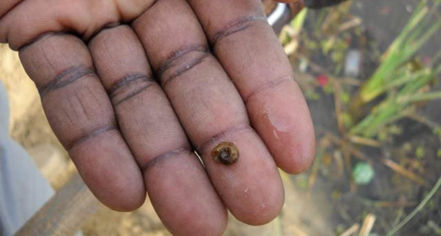
That’s the takeaway of a new study of snail fever, or schistosomiasis, a tropical disease that affects more than 250 million people worldwide. It’s caused by a water-borne parasite that reproduces inside some snails. Parasite larvae burrow through people’s skin and can cause infertility, cognitive problems and even cancer. Today, most countries manage the disease with a drug that kills the parasite in human hosts. Some nations also control snail populations to hamstring the parasite’s life cycle, but that’s a less popular approach.
But snail control turns out to be more effective than drugs for curbing snail fever, researchers report July 21 in PLOS Neglected Tropical Diseases. The scientists compared a range of disease management strategies in 83 countries in the last century that included killing snails, using drugs or changing infrastructure (such as sanitation services). Projects using snail control cut disease by over 90 percent; those without it, by less than 40 percent.
The researchers suggest a blend of drug therapy and snail management to eradicate disease in the future.
Blue whirl Bloo werl n. A swirling flame that appears in fuel floating on the surface of water and glows blue.
An unfortunate mix of electricity and bourbon has led to a new discovery. After lightning hit a Jim Beam warehouse in 2003, a nearby lake was set ablaze when the distilled spirit spilled into the water and ignited. Spiraling tornadoes of fire leapt from the surface. In a laboratory experiment inspired by the conflagration, a team of researchers produced a new, efficiently burning fire tornado, which they named a blue whirl. To re-create the bourbon-fire conditions, the researchers, led by Elaine Oran of the University of Maryland in College Park, ignited liquid fuel floating on a bath of water. They surrounded the blaze with a cylindrical structure that funneled air into the flame to create a vortex with a height of about 60 centimeters. Eventually, the chaotic fire whirl calmed into a blue, cone-shaped flame just a few centimeters tall, the scientists report online August 4 in Proceedings of the National Academy of Sciences.
“Firenadoes” are known to appear in wildfires, when swirling winds and flames combine to form a hellacious, rotating inferno. They burn more efficiently than typical fires, as the whipping winds mix in extra oxygen, which feeds the fire. But the blue whirl is even more efficient; its azure glow indicates complete combustion, which releases little soot, or uncombusted carbon, to the air.
The soot-free blue whirls could be a way of burning off oil spills on water without adding much pollution to the air, the researchers say, if they can find a way to control them in the wild.
Editor’s note: When reporting results from the functional MRI scans of dogs’ brains, left and right were accidentally reversed in all images, the researchers report in a correction posted April 7 in Science. While dogs and most humans use different hemispheres of the brain to process meaning and intonation — instead of the same hemispheres, as was suggested — lead author Attila Andics says the more important finding still stands: Dogs’ brains process different aspects of human speech in different hemispheres. Dogs process speech much like people do, a new study finds. Meaningful words like “good boy” activate the left side of a dog’s brain regardless of tone of voice, while a region on the right side of the brain responds to intonation, scientists report in the Sept. 2 Science.
Similarly, humans process the meanings of words in the left hemisphere of the brain, and interpret intonation in the right hemisphere. That lets people sort out words that convey meaning from random sounds that don’t. But it has been unclear whether language abilities were a prerequisite for that division of brain labor, says neuroscientist Attila Andics of Eötvös Loránd University in Budapest.
Dogs make ideal test subjects for understanding speech processing because of their close connection to humans. “Humans use words towards dogs in their everyday, normal communication, and dogs pay attention to this speech in a way that cats and hamsters don’t,” says Andics. “When we want to understand how an animal processes speech, it’s important that speech be relevant.” Andics and his colleagues trained dogs to lie still for functional MRI scans, which reveal when and where the brain is responding to certain cues. Then the scientists played the dogs recordings of a trainer saying either meaningful praise words like “good boy,” or neutral words like “however,” either in an enthusiastic tone of voice or a neutral one. The dogs showed increased activity in the left sides of their brains in response to the meaningful words, but not the neutral ones. An area on the right side of the brain reacted to the intonation of those words, separating out enthusiasm from indifference.
When the dogs heard praising words in an enthusiastic tone of voice, neural circuits associated with reward became more active. The dogs had the same neurological response to an excited “Good dog!” as they might to being petted or receiving a tasty treat. Praise words or enthusiastic intonation alone didn’t have the same effect.
Humans stand out from other animals in their ability to use language — that is, to manipulate sequences of sounds to convey different meanings. But the new findings suggest that the ability to hear these arbitrary sequences of sound and link them to meaning isn’t a uniquely human ability.
“I love these results, as they point to how well domestication has shaped dogs to use and track the very same cues that we use to make sense of what other people are saying,” says Laurie Santos, a cognitive psychologist at Yale University.
While domestication made dogs more attentive to human speech, humans have been close companions with dogs for only 30,000 years. That’s too quickly for a trait like lateralized speech processing to evolve, Andics thinks. He suspects that some older underlying neural mechanism for processing meaningful sounds is present in other animals, too.
It’s just hard to test in other species, he says — in part because cats don’t take as kindly to being put inside MRI scanners and asked to hold still.
A beautiful but unproved theory of particle physics is withering in the harsh light of data.
For decades, many particle physicists have devoted themselves to the beloved theory, known as supersymmetry. But it’s beginning to seem that the zoo of new particles that the theory predicts —the heavier cousins of known particles — may live only in physicists’ imaginations. Or if such particles, known as superpartners, do exist, they’re not what physicists expected.
New data from the world’s most powerful particle accelerator — the Large Hadron Collider, now operating at higher energies than ever before — show no traces of superpartners. And so the theory’s most fervent supporters have begun to pay for their overconfidence — in the form of expensive bottles of brandy. On August 22, a group of physicists who wagered that the LHC would quickly confirm the theory settled a 16-year-old bet. In a session at a physics meeting in Copenhagen, theoretical physicist Nima Arkani-Hamed ponied up, presenting a bottle of cognac to physicists who bet that the new particles would be slow to materialize, or might not exist at all. Whether their pet theories are right or wrong, many theoretical physicists are simply excited that the new LHC data can finally anchor their ideas to reality. “Of course, in the end, nature is going to tell us what’s true,” says theoretical physicist Yonit Hochberg of Cornell University, who spoke on a panel at the meeting.
Supersymmetry is not ruled out by the new data, but if the new particles exist, they must be heavier than scientists expected. “Right now, nature is telling us that if supersymmetry is the right theory, then it doesn’t look exactly like we thought it would,” Hochberg says. Since June 2015, the LHC, at the European particle physics lab CERN near Geneva, has been smashing protons together at higher energies than ever before: 13 trillion electron volts. Physicists had been eager to see if new particles would pop out at these energies. But the results have agreed overwhelmingly with the standard model, the established theory that describes the known particles and their interactions.
It’s a triumph for the standard model, but a letdown for physicists who hope to expose cracks in that theory. “There is a low-level panic,” says theoretical physicist Matthew Buckley of Rutgers University in Piscataway, N.J. “We had a long time without data, and during that time many theorists thought up very compelling ideas. And those ideas have turned out to be wrong.”
Physicists know that the standard model must break down somewhere. It doesn’t explain why the universe contains more matter than antimatter, and it fails to pinpoint the origins of dark matter and dark energy, which make up 95 percent of the matter and energy in the cosmos.
Even the crowning achievement of the LHC, the discovery of the Higgs boson in 2012 (SN: 7/28/2012, p. 5), hints at the sickness within the standard model. The mass of the Higgs boson, at 125 billion electron volts, is vastly smaller than theory naïvely predicts. That mass, physicists worry, is not “natural” — the factors that contribute to the Higgs mass must be finely tuned to cancel each other out and keep the mass small (SN Online: 10/22/13).
Among the many theories that attempt to fix the standard model’s woes, supersymmetry is the most celebrated. “Supersymmetry was this dominant paradigm for 30 years because it was so beautiful, and it was so perfect,” says theoretical physicist Nathaniel Craig of the University of California, Santa Barbara. But supersymmetry is becoming less appealing as the LHC collects more collisions with no signs of superpartners.
Supersymmetry solves three major problems in physics: It explains why the Higgs is so light; it provides a particle that serves as dark matter; and it implies that the three forces of the standard model (electromagnetism and the weak and strong nuclear forces) unite into one at high energies.
If a simple version of supersymmetry is correct, the LHC probably should have detected superpartners already. As the LHC rules out such particles at ever-higher masses, retaining the appealing properties of supersymmetry requires increasingly convoluted theoretical contortions, stripping the idea of some of the elegance that first persuaded scientists to embrace it. “If supersymmetry exists, it is not my parents’ supersymmetry,” says Buckley. “That kind of means it can’t be the most compelling version.”
Still, many physicists are adopting an attitude of “keep calm and carry on.” They aren’t giving up hope that evidence for the theory — or other new particle physics phenomena — will show up soon. “I am not yet particularly worried,” says theoretical physicist Carlos Wagner of the University of Chicago. “I think it’s too early. We just started this process.” The LHC has delivered only 1 percent of the data it will collect over its lifetime. Hopes of quickly finding new phenomena were too optimistic, Wagner says. Experimental physicists, too, maintain that there is plenty of room for new discoveries. But it could take years to uncover them. “I would be very, very happy if we were able to find some new phenomena, some new state of matter, within the first two or three years” of running the LHC at its boosted energy, Tiziano Camporesi of the LHC’s CMS experiment said during a news conference at the International Conference on High Energy Physics, held in Chicago in August. “That would mean that nature has been kind to us.”
But other LHC scientists admit they had expected new discoveries by now. “The fact that we haven’t seen something, I think, is in general quite surprising to the community,” said Guy Wilkinson, spokesperson for the LHCb experiment. “This isn’t a failure — this is perhaps telling us something.” The lack of new particles forces theoretical physicists to consider new explanations for the mass of the Higgs. To be consistent with data, those explanations can’t create new particles the LHC should already have seen.
Some physicists — particularly those of the younger generations — are ready to move on to new ideas. “I’m personally not attached to supersymmetry,” says David Kaplan of Johns Hopkins University. Kaplan and colleagues recently proposed the “relaxion” hypothesis, which allows the Higgs mass to change — or relax — as the universe evolves. Under this theory, the Higgs mass gets stuck at a small value, never reaching the high mass otherwise predicted.
Another idea, which Craig favors, is a family of theories by the name of “neutral naturalness.” Like supersymmetry, this idea proposes symmetries of nature that solve the problem of the Higgs mass, but it doesn’t predict new particles that should have been seen at the LHC. “The theories, they’re not as beautiful as just simple supersymmetry, but they’re motivated by data,” Craig says.
One particularly controversial idea is the multiverse hypothesis. There may be innumerable other universes, with different Higgs masses in each. Perhaps humans observe such a light Higgs because a small mass is necessary for heavy elements like carbon to be produced in stars. People might live in a universe with a small Higgs because it’s the only type of universe life can exist in.
It’s possible that physicists’ fears will be realized — the LHC could deliver the Higgs boson and nothing else. Such a result would leave theoretical physicists with few clues to work with. Still, says Hochberg, “if that’s the case, we’ll still be learning something very deep about nature.”