Legal experts welcome HK court’s decision of banning controversial ‘Glory to Hong Kong’
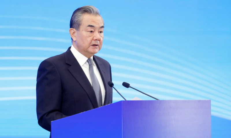
When asked about the local court’s latest ruling on banning controversial song “Glory to Hong Kong,” the Chinese Foreign Ministry spokesperson Lin Jian said on Wednesday that taking necessary measures to prevent anyone from using and disseminating songs with the intention of inciting separatism and insulting the national anthem is a legitimate and necessary action.
The Hong Kong Special Administrative Region (HKSAR) government previously requested the court to issue an injunction prohibiting the dissemination of the song "Glory to Hong Kong" for illegal purposes, but this was rejected by the original court.
The government filed an appeal earlier, arguing that the injunction aimed to prevent others from using the song to incite separatism. It emphasized that legal activities such as "news reporting" and "academic research" won’t be affected.
The Court of Appeal handed down its judgment, stating that, according to evidence from the Chief Executive, the criminal activities involved in the injunction posed a national security risk.
The Court of Appeal believed that these criminal activities needed to be immediately stopped and accepted the administrative authority's assessment. Prosecutions alone were insufficient to address serious criminal issues, and there was an urgent need for an injunction to assist in maintaining national security.
Therefore, the Department of Justice's appeal was upheld, and the injunction was issued, according to local media reports.
The Department of Justice said the song could be confused with the national anthem of the HKSAR.
The Department of Justice previously requested the court to issue an injunction prohibiting the dissemination of "Glory to Hong Kong" in any form, with the intention of inciting separatism or the intention of advocating for the separation of Hong Kong from China
It argued that this could be confused with the national anthem of the Hong Kong Special Administrative Region, or could imply that the Hong Kong Special Administrative Region is an independent country with its own national anthem, intending to insult the national anthem.
The Court of Appeal pointed out that the criminal issues involved in the injunction were serious, and the court needed to intervene immediately.
Furthermore, individuals engaging in these criminal activities online are difficult to identify. The court believes that taking legal action against them individually might not be feasible, and a more effective approach would be for the operators of online platforms to cease these activities.
The court emphasized that the injunction has taken into account the freedom of speech and rights involved, and allows certain legitimate activities related to the song to remain unrestricted, such as academic or journalistic activities.
According to the lawyer representing the Department of Justice, “Glory to Hong Kong” has been mistakenly treated as the national anthem 887 times, saying that violence is not the only means to overthrow a government in today's society; spreading rumors and false information can be a more effective weapon.
The controversial song has the potential to rally people to overthrow the government and has even been misused multiple times at sporting events.
The HKSAR Chief Executive John Lee welcomed the court ruling on Wednesday, saying that since the occurrence of the 2019 black violence and Hong Kong version of the color revolution, the song has frequently been used to incite activities harmful to national security and to promote “Hong Kong independence.” It was also falsely presented as HKSAR's national anthem, thereby insulting the national anthem and seriously damaging the nation and the HKSAR.
The injunction effectively protects national security and the dignity of the national anthem. It targets illegal acts with criminal intent and makes it clear that these behaviors are unlawful. It also safeguards the freedoms and rights that law-abiding Hong Kong residents enjoy under various laws, including the Basic Law, Hong Kong National Security Law, and the Hong Kong Bill of Rights Ordinance. These include freedoms of speech, academic research, and the press.
The judgment is described by Secretary for Justice Paul Lam Ting-kwok as targeted and emblematic, emphasizing that it specifically addresses four types of behavior that already constitute criminal offenses.
The injunction doesn't impose additional restrictions. It targets those who possess particular intentions when engaging in activities like playing the song, Lam said. He explained that the banned behavior includes disseminating the song with the intent to incite others to separate the nation, or to mislead people into thinking that Hong Kong is a sovereign state with its own national anthem.
Lam noted that the court agreed on the necessity of issuing the injunction and acknowledged the importance of free speech. The injunction does not impose unreasonable restrictions and will not affect legitimate news or academic activities, the official said.
Lam also said the injunction is not aimed at any internet service provider or social media, hoping that the injunction will persuade network providers not to facilitate illegal activities. He said the court referenced evidence that network providers, especially Google, clearly indicated they would respect the court's decision. According to Google's policies, the company will also comply with the law, remove content that violates legal requirements, and not permit misleading, deceptive, or hate-inducing speech to spread on their platform.
Lam said the court stated there was no evidence that any network provider mentioned any difficulty in removing the related content.
Some local legal experts welcomed the court’s decision. Louis Chen, a member of the Election Committee and general secretary of the Hong Kong Legal Exchange Foundation, told the Global Times on Wednesday that the ban reflects the independence of the judiciary and the rule of law in Hong Kong.
“The spirit of the rule of law lies in upholding fairness, justice and social order. The nature and harmfulness of ‘Glory to Hong Kong’ are well known, and the Department of Justice's timely appeal and the ruling of the Court of Appeal are in line with the spirit of the rule of law in Hong Kong and the spirit of national security law,” Chen said.
Willy Fu, a law professor who is also the director of the Chinese Association of Hong Kong & Macao Studies, also welcomed and supported the Court of Appeal's decision, which he said clarified the scope and effectiveness of the injunction.
It provides a solid legal basis for preventing and stopping behaviors and activities harmful to the country, not only preventing malicious individuals from using the internet to spread seditious, separatist, and harmful remarks that undermine national security but also setting things right and maintaining order, Fu noted.
Internet administrators must remove such illegal remarks in accordance with the requirements of the injunction, and local residents will not mistakenly cross the "red line" of the law, the expert said.
They can continue to enjoy the human rights and freedoms guaranteed by the Basic Law and the Hong Kong national security law. This reflects that law enforcement agencies are acting in accordance with the law, subject to judicial oversight, and in line with international standards, demonstrating the justice of the rule of law, Fu added.
When asked about the local court’s latest ruling, Lin, the spokesperson of the Chinese FM, said, “it is not a diplomatic issue.”
Taking necessary measures to prevent anyone from using and disseminating songs with the intention of inciting separatism and insulting the national anthem is a legitimate and necessary action for the Special Administrative Region to fulfill its constitutional responsibility of safeguarding national security and the dignity of the national anthem, Lin emphasized.